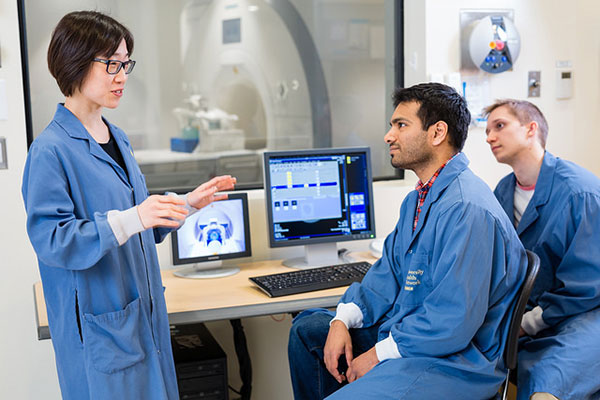
Last summer, Hai-Ling Margaret Cheng and her team of biomedical engineering researchers developed a new way to monitor the effectiveness of stem cell therapy for heart failure patients. Now, she’s teaching others the techniques, and how they can be used to create new medical diagnostic approaches.
“Magnetic resonance imaging (MRI) is a powerful tool that can offer a wealth of information about the human body,” said Cheng, a MRI expert and a professor of biomedical and electrical engineering at the University of Toronto. “It has the capability to provide a lot more than a pretty picture. Understanding its broad range of capabilities and some of the latest imaging techniques can help medical professionals, engineers and scientists come up with new solutions to address pressing human health challenges.”
Cheng’s new course, BME 1466—Advanced Topics in Magnetic Resonance Imaging, will be offered to graduate students at U of T starting in January 2018. Some of the topics she’ll cover already have a major impact on how scientists and engineers approach several challenging diseases.
“For example, we’re going to look at how to capture precise and useful images of blockage in the heart’s smallest arteries, and how certain drugs might pass through the blood-brain barrier for cancer treatment,” said Cheng. “We’re also going to study some of the current obstacles to diagnosis and treatment in relation to MRI techniques.”
For some current graduate students, this course has the potential to greatly accelerate their research.
“This course will give me the tools to understand the bigger picture behind my research where I focus a lot on MRI acquisition techniques for diagnosis purposes,” said Eric Zakher, an electrical engineering master’s student. “MRI is one of the most complex imaging modalities and this course is a great opportunity to get exposure to a field that is revolutionizing the diagnosis and treatments of many medical conditions.”
“I was taught computed tomography and nuclear medicine techniques during my clinical training so being able to learn about MRI has increased my attraction to this field,” said Tameshwar Ganesh, a pharmaceutical sciences PhD student and a registered nuclear medicine technologist. “This course will help me with my doctoral thesis where I will use MRI to evaluate vascular health.”
The availability of BME 1466 also adds to the Institute of Biomaterials & Biomedical Engineering’s (IBBME) course repertoire behind its Nanotechnology, Molecular Imaging & Systems Biology research theme.
“There are really no comparable courses offered in this area,” said Professor Christopher Yip, IBBME’s former director and U of T’s associate vice-president of international partnerships.
“What is really compelling will be how she will be bringing current, leading-edge research in MRI into her lectures to engage students — helping showcase how this tool can provide insights into molecular and cellular dynamics, and physiological state. In essence — truly functional imaging.”