
The University of Toronto is home to many experts who study how cities can be improved. One aspect of cities that may be taken for granted is one of the most important: water supply.
At U of T, water conservation efforts have been underway since the 1970s. For example, underground cisterns on the downtown campus collect rainwater, which is then used by a smart irrigation system that only waters lawns if there is no rain in the forecast. But as cities continue to grow, so does the need for everyone to protect and manage water resources.
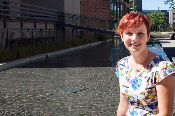
Enter Professor Jennifer Drake (CivE), an expert in water security.
Drake’s research expertise includes stormwater systems, watershed planning and stormwater management. She’s especially passionate about building and managing urban water systems that minimize the impact on the natural environment.
The increasing frequency of extreme weather events such as droughts and floods is drawing attention to Drake’s research and the challenges and opportunities of protecting urban water resources in the 21st century.
U of T writer Dominic Ali spoke with Drake about the importance of water for cities in the 21st century.
Why is water management so important?
We only have a finite amount of water and it is cycled in the environment over and over again. This means that no matter where you are, the water in the natural environment will be used by someone for drinking, irrigation, recreation, etc. Water security includes issues of availability and water quality.
My research focuses on issues of quantity such as flooding (i.e., protecting us from when there is too much water) and quality such as developing new technologies to improve the quantity of urban runoff before it is returned to a natural system like a creek or lake.
Canada’s infrastructure deficit is estimated at $123 billion of which $31 billion is for water/wastewater. Improvements to water infrastructure are critical to the Canadian economy. Moreover, when we invest in our water resources and foster healthy aquatic environments we make our cities more liveable and create opportunities for residents to experience the beauty and peace of the natural environment.
Why is low-impact development seen as better for water management?
Our water resources are much more resilient and secure when we work with nature instead of against it. For example, if you live in the U.S. southwest, a region of water scarcity, your lawn should be landscaped with drought-resistant plants, not turf grass. In Ontario, low-impact development practices aim to restore the hydrology that is often lost as a result of urbanization. This is achieved through innovative technologies like green roofs, bioretention systems and permeable pavements.
What are some of the water challenges faced by cities?
One of the biggest challenges facing cities like Toronto is the uncertainty regarding water availability in the future. All of our economic and social systems depend on reliable source waters (groundwater, lakes, rivers and streams) with sufficient quantity and quality. Not only do we require water for drinking and recreation but all agricultural, manufacturing and resource-based industries require secure water supplies, too.
Climate change will alter the availability of water at local and national levels. Cities need to invest in infrastructure, which improves our resilience during extreme weather. In Ontario it is anticipated that climate change will change the type (snow vs. rain) and timing of precipitation. To adapt to climate change infrastructure investment, replacement and maintenance will be essential for cities. Some cities, such as Kitchener and Mississauga, are already adapting by developing new revenue mechanisms to support these costs.
With a Toronto election coming up, what question would you pose to the candidates about water security?
Toronto has implemented some very progressive programs such as the Green Roof Bylaw and the Mandatory Downspout Disconnection Program that are aimed at reducing flooding and improving water quality. However, despite these actions, flooding continues to affect Torontonians quite regularly.
From the mayoral candidates I would like to know what programs they would advance to continue reducing the occurrence of flooding within the city and minimizing the impact of flooding on Torontonians.
What first attracted you to this field?
I first became interested in urban water management and water security as an undergraduate student during a summer co-op placement with the City of Burlington. I observed first-hand the challenges associated with managing water infrastructure. This inspired me to ultimately research water security.
One of my tasks was to research maintenance costs for stormwater management pond cleaning. I was amazed to discover that, at the time, Ontario’s municipalities had invested in ponds for flood and water quality control but did not have sufficient funds to conduct the cleaning projects that need to be completed every 10 to 15 years.
This issue became even more compelling when I realized that a mid-sized Ontario municipality may operate 50 to 100 ponds and a single cleaning project could cost over $500,000.
Are any cities doing innovative things to improve water security?
There are a surprising number of success stories. Las Vegas, a city more famous for excess than conservation, is a great example. While the population of Las Vegas has tripled over the last 20 years, its per capita water use has dropped by 31 per cent. This means that the total water used today is almost the same as it was 20 years ago. Las Vegas has achieved this through changing water rates, supporting conservation and investing the infrastructure to allow for water re-use.

According to Shums Kassam (EngSci 1T5), a U of T Engineering degree equips students with many valuable things, but a solid fashion sense isn’t necessarily one of them.
Two things, among others, that Kassam claims he has gained from his engineering degree are both the competencies to build a digital personal stylist app called Blynk, as well as the connections that inspired him to launch it into a business, which is already seeing success on the market.
Kassam and fellow Blynk co-founder Jaclyn Ling designed their business around an app that combines user style preferences, instant purchases and the addictive swipe-right, swipe-left format.
“Through a Tinder-like interface, users swipe through inspiration photos – left to dislike, right to like,” said Kassam. “Blynk learns users’ style preferences and recommends a full outfit that users can buy based on what they like. Users also have the ability to build and share outfits, and even request outfits from fashion stylists – all for free.”
The duo developed Blynk both through U of T Engineering’s Entrepreneurship Hatchery and as part of the Next 36 entrepreneurship development program, co-founded by U of T’s Ajay Agrawal.
Since going through the Next 36, Kassam and Ling launched Blynk to great market and media interest, including features in Toronto Life, Global TV, the Globe and Mail and more. Kassam says the attention has been driven largely by the company’s entrepreneurial drive, as they’ve already pitched at industry showcases and even won a $100,000 investment for top spot in the International Startup Festival competition.
“Blynk’s vision is to provide free and accessible fashion advice to everyone,” he said, “through a simple, fun and addicting platform. An average personal stylist costs over $100 an hour. Blynk replicates this value for free, anytime and anywhere.”
Kassam spoke with U of T News about what’s next for the growing company.
It all started with a wardrobe problem
SK: Jaclyn and I arrived at this startup idea when she took me shopping early in December and gave me style suggestions. My confidence improved, and I really liked dressing better. We started working on Blynk in early April – developing it on Android while finishing up exams at school. Since then the startup has been going well. We won $100,000 from the International Startup festival and have had over 8,000 downloads while the app has been only out for about two months.
An app empowering fashion fans and fashion-challenged alike
SK: Blynk is for a variety of people – for someone like myself, I use Blynk because it helps me find products that I like and helps me define my own style. However, Blynk is also for fashion enthusiasts who want inspiration, or to share their love of fashion with others by posting content. Essentially, Blynk is for those who want to improve their style or those who enjoy expressing their style.
Blynk taps into the fashion blog community
SK: The fashion photos [featured on Blynk] come from different bloggers. We have partnered with a few fashion posts to upload content. In a newer version of Blynk [coming out within the next few weeks], users can upload their own content and, if approved, the content will be put in the feed. We are also planning, and already are starting, to partner with professional stylists. We continue to approve more stylists to our app.
Profits driven by users and vendors
SK: Many retail stores have contacted us because of interest in native advertising and in affiliate programs, where we can make a commission.
The real reason a fashion-challenged engineer devotes his time to a styling app
SK: I have a real passion for entrepreneurship – I see so much value in it, because it is a way to provide jobs and improve economic growth, while also trying to disrupt systems.
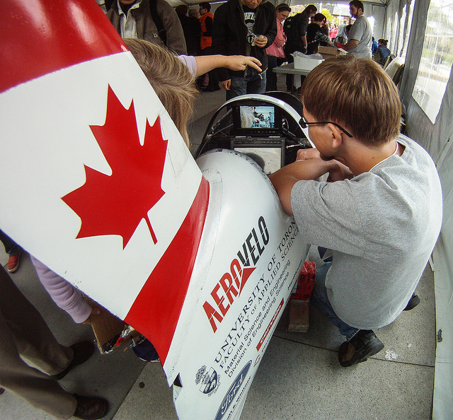
For anyone who makes anything – from R2D2 robots to DIY skateboards to 3D-printed chocolate sculptures and beyond – participating in the Maker Faire means entering into a worldwide family-friendly carnival of wondrous, inventive physical stuff.
Launched as a showcase of cool inventions from California’s Bay area in 2006, satellite Maker Faires have since popped up around the globe in locations like Kenya, Norway and Italy, with a second annual instalment hosting thousands of ‘makers’ in Toronto on November 22 and 23.
“We are looking forward to getting feedback from the community as well as seeing what’s out there from other local startups and makers,” said Michael Helander (EngSci 0T7, MSE PhD 1T2), co-founder of OTI Lumionics, a startup founded on U of T Engineering research innovations in organic LED lighting. “We strongly believe in the importance of building the local startup community and so we are delighted to be attending the Maker Faire in Toronto.”
Maker Faire debuted in Toronto last fall, gathering 4,000 makers of all ages and experience levels as they shared and experimented with a wild range of projects. Now, the Toronto team is raising the stakes, as they are set to present more than 100 exhibits – including the OTI Lumionics demo for their aerelight lamps – as well as a range of workshops and events in the Toronto Reference Library.
“Maker Faire is a unique opportunity to showcase inventions, meet fellow tinkerers and see the diverse maker crowd response to projects,” said Elena Yunusov, a co-organizer of Toronto Maker Faire and a digital communications strategist at U of T’s Faculty of Medicine.
“It’s important to give students an opportunity to play with technology, realize that everyone can make things and see that tinkering is a vital aspect of innovation and learning.”
Last year’s Faire drew U of T startups and students as attendees and exhibitors. Now, the call is for more university makers and audiences to join for the 2014 edition: the Toronto Maker Faire is accepting exhibitor applications until September 21, and offers free tickets for attendees online.
Yunusov offers three reasons U of T makers should submit their ideas for showcase, discussion and play at Toronto Maker Faire 2014:
1. Put your early-stage project into the hands of real users of all ages
EY: Last year, a startup named Battlegrounds put on an advanced laser tag game. After two years of design and testing, this was their first public demonstration and hundreds of kids got to play it. When a couple of their 3D-printed laser tag guns broke, the kids using them were upset they had broken something. The makers, on the other hand, were super happy because they finally knew what they needed to fix to make the equipment more durable. It was awesome because the kids were super surprised the makers were giving them bear hugs and gifts for breaking things!
2. Experience ideas of the future with your own eyes – and hands
EY: The Maker Faire is a great way to get inspired by all the amazing inventions that exist and to see it all first-hand – get a glimpse of the kind of future that is unevenly distributed.”
3. Build a business grounded in imagination
EY: The Maker Faire attracts entrepreneurs because they can showcase and prototype their inventions, develop new ideas, gauge public reaction and chat with other entrepreneurs. Many of the projects are crowdfunded, which is amazing to see.

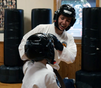
First-year engineering student Quinton Lowe (EngSci 1T8) holds a black belt in karate, is a political advocate and is passionate about engineering. Now, he’s also one of two U of T students to receive the Schulich Leaders Scholarship, a prestigious $80,000 award started by business mogul and philanthropist Seymour Schulich.
Supporting 40 students annually across Canada, the scholarship rewards students pursuing undergraduate degrees in STEM (science, technology, engineering and math) who have demonstrated significant leadership qualities.
“Congratulations to Quinton Lowe for winning this prestigious award,” said Micah Stickel, Chair, First Year Engineering. “This recognition is a testament to the innovative thinkers and inspirational leaders our engineering programs attract.”
U of T’s Xarissa Thompson spoke with Lowe about the award and why he chose to study engineering at the University of Toronto.
What drew you to the University of Toronto – and why STEM?
I applied to the University of Toronto because I want the challenge and excitement of studying at the best engineering school in Canada. Top employers recognize the value of a U of T degree and I’ve heard news stories about the inventions and discoveries being made at this university. I also love Toronto and liked the idea of [living] downtown, where I could explore new things and have new experiences.
My interest in STEM courses really began in grade 11, when my courses started to focus on the specific sciences. I took a physics course that I really enjoyed and started to feel a pull towards a career in science.
 How important are co-curricular activities and volunteer work?
I am proud to have earned a black belt in karate. It took many years of hard work and dedication. A black belt’s duty is to help others learn and master the art, so I have assisted a weekly class where I led the warm-up and basics and individually assisted students when they were struggling.
My passion for politics began when I was selected to serve a term of duty as a legislative page. I [stayed with] an aunt who lived in Toronto and I spent a month of the school year at Queen’s Park. We would fetch glasses of water, make photocopies or run messages for the MPPs. I hadn’t known much about how politics worked previously and this gave me a first-hand view of the process.
At age 14, I decided to get involved with the local riding associations and volunteered hundreds of hours in preparation for both federal and provincial elections. I found it very rewarding serving the community through canvassing, helping disabled and elderly citizens, and acting as an election scrutineer. I have remained heavily involved in politics and am the youngest person ever to be elected to both the federal and provincial conservative riding associations’ board of directors.
Any plans for the future?
An engineering degree is the best possible undergraduate degree for someone with my interests to obtain. Ultimately I would like to earn a graduate degree. I may end up working in the engineering field, but law and politics are also possibilities.
Learn more about the Schulich Leader Scholarships.

Professor Milica Radisic (IBBME, ChemE) is among three U of T researchers named to the inaugural cohort of the Royal Society of Canada’s new College of New Scholars, Artists and Scientists – an initiative that recognizes the emerging generation of Canadian intellectual leaders.
Radisic, a researcher in both the Institute for Biomaterials and Biomedical Engineering (IBBME) and the Department of Chemical Engineering and Applied Chemistry (ChemE) at U of T, is a world leader in the field of cardiovascular tissue engineering. She was cited by the RSC for her innovative techniques in designing and developing new heart tissue derived from stem cells. Her contributions have been recognized by multiple national and international prizes, most recently an NSERC Steacie Fellowship.
“Milica Radisic has made exceptional contributions in the fields of tissue engineering and regenerative medicine” said Dean Cristina Amon. “Her pioneering research has the potential to revolutionize the treatment of cardiovascular disease. She is most deserving of this prestigious new honour from the Royal Society of Canada.”
Radisic joins three other U of T researchers in the College, including religious studies professor Amira Mittermaier and historian Nathalie E. Rothman. In total, the RSC has named 91 members to the College from 52 Canadian universities and other institutions.
The College of New Scholars, Artists and Scientists was created to gather scholars, artists and scientists at a highly productive stage of their careers into a single collegium where new advances in understanding will emerge from the interaction of diverse intellectual, cultural and social perspectives.
The initiative’s mandate is to address issues of particular concern to the group of interdisciplinary collaborators, for the advancement of understanding and the benefit of society, taking advantage of the interdisciplinary approaches fostered by the RSC.
Learn more about RSC’s College of New Scholars, Artists and Scientists.

What do soap bubbles and grasshoppers have in common? As it turns out, they can both be used to help students learn about modern materials and their engineering applications.
This summer, Dr. Scott Ramsay and Professor Uwe Erb (both MSE) each led an undergraduate student team from the Department of Materials Science & Engineering (MSE). Students teamed up to develop new educational kits that will help fellow students learn introductory engineering concepts.
The first group worked with Ramsay on a project called Materials One as part of the engineering course, MSE101. They collaborated on new methods to help first-year students grasp challenging concepts in materials science. The other group, part of the nanOntario outreach program led by Erb, developed instructional kits for high school students.
“The concepts we teach in materials engineering are often new to our students and can be challenging to grasp,” said Ramsay. “There isn’t really an equivalent prerequisite course at the Ontario secondary school level. That’s why we have students come up with educational aids – to facilitate learning at a level that speaks to other students. And, it’s really a lot of fun for everyone.”
Take a look at the two groups that came up with fun – and sometimes sticky – learning activities.
Materials One

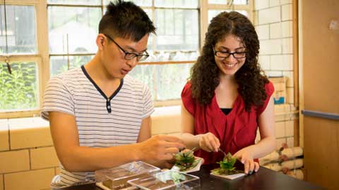
Undergraduate students Anastasia Alksnis (MSE 1T6), Roberto Aurilio (MSE 1T5), Daniel Levitt (MSE 1T7), and Xiaoji (Iris) Zhang (MSE 1T5) developed a series of hands-on activities and learning aids that complement MSE 101 – the first-year materials science and engineering course offered across the Faculty.
One of the activities the group came up with asks students to blow soap bubbles onto a flat surface forming a so-called ‘bubble raft.’ Developed in the late 1940s, this technique was first used by researchers to model atoms in solids. Now reimagined for MSE 101, students will be challenged to take photos of their bubble rafts, which show the best examples of various microstructural features – features that contribute to materials properties and performance.
“Working on the Materials One project was a great way for me to reinforce what I had learned in MSE 101,” said Levitt, whose group was supported by the Faculty’s Engineering Instructional Innovation Program. “I hope our efforts will help future students engage with the curriculum and build a solid foundation for their upper year studies.”
Watch Dr. Scott Ramsay perform the elephant toothpaste experiment at a local elementary school, illustrating catalyzed decomposition of hydrogen peroxide – an experiment prepared by Materials One project student Xiaoji (Iris) Zhang.
nanOntario
Now in its fifth year, nanOntario is a youth outreach program led by Professor Uwe Erb (MSE) aimed at educating secondary school students about bio-inspired nanotechnology found in Ontario’s outdoors.
Tasked with designing instructional kits to facilitate the course, undergraduates must think deeply about materials science subjects in order to best deliver the information. In this way, the learning is two-fold, as both high school and university-level participants benefit from the course.
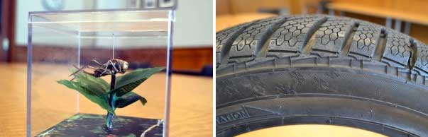
This year’s theme was “Hexagonal Structures”. Participants investigated the prevalence of six-sided surface structures in nature, and how humans have mimicked these shapes to enhance technologies.
For example, katydids – an insect similar to the grasshopper – have hexagonal micro-patterns on their legs that help them attach to surfaces when jumping from place to place. A similar pattern can be found in certain winter tires to improve traction for driving safely in slippery conditions.
Partnered with Hitachi High-Technologies Canada (HHTC), participants of the course have access to a portable scanning electron microscope that enable discovery at the atomic level, including a close-up look at the surface structure of insect legs.
“As students shift more of their studies towards the use of online materials, one of the best functions of classroom and tutorial time is to make use of demonstrations and hands on activities to facilitate different modes of learning,” said Professor Jun Nogami, Chair of the Department of Materials Science & Engineering. “The ideal way to develop these new teaching materials is to involve students in their development from the very beginning.”