Microscopes are some of the most powerful tools in cell biology — but what if the cell component that needs to be imaged is smaller than the wavelengths of visible light? A new study from Professor Chris Yip (ChemE, BME) proposes a solution, one that could help advance research into cancer and other diseases.
“There are a couple of existing techniques to image things that are smaller than light, but they have drawbacks,” says Yip.
“One is electron microscopy, which relies on firing electrons at a sample. Another is atomic force microscopy, which involves dragging a tiny needle tip across a surface. But if the feature you’re trying to image doesn’t have a distinct shape or a unique electron density, it’s hard to identify what you’re looking at. And in our case, the thing keeps moving around.”
The challenge facing Yip and his collaborators — including Professor Scott Gray-Owen (Molecular Genetics, Medicine) and BME PhD candidate Amine Driouchi — was to study how a particular protein known as CEACAM1 distributes itself over the surface of a living cell in real time.
Cell surface proteins are key to many different biological processes, influencing how cells take in nutrients and how they send signals to one another. Viruses also use cell surface proteins to get inside cells and hijack their biochemical machinery in order to replicate.
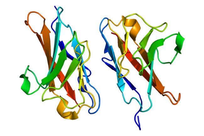
CEACAM1 is a cell surface protein found on certain types of cancer cells, and is suspected of playing a role in cell adhesion within tumours. How exactly it does this is not well understood, but the team believes that it may have something to do with the way it is arranged.
“On the surface of the cell, CEACAM1 can exist as a monomer — that is, by itself — or as a dimer, meaning two of them stuck together,” says Yip. “It can also form aggregates or clusters. What we wanted to see was whether these different arrangements had any impact on its function in terms of helping cancer cells adhere to each other.”
In research recently published in the Journal of Biological Chemistry, the team applied a variation of a technique known as Förster resonance energy transfer (FRET) to see whether CEACAM1 was arranged in clusters, dimers or individual molecules.
Conventional FRET is used to tell whether two different proteins are interacting with each other. Both proteins are tagged with unique fluorescent marker molecule, each of which absorbs light at one wavelength and emits it at another. Crucially, the emission wavelength of marker A is the same as the excitation wavelength of marker B.
If the proteins are close together — which suggests they are interacting — exciting marker A will cause energy to jump across and light up marker B. If only marker A lights up, it means the proteins are too far apart to make this jump.
But in this case, the team was dealing with a single protein interacting with copies of itself, so it could not be tagged with two different markers. To overcome this challenge, the team made use of a different feature of light: its polarization.
After all the CEACAM1 molecules were tagged with the same fluorescent marker, the team hit the sample with polarized light at the right wavelength to excite this marker. When the fluorescent marker got excited and released a photon in response, the team tracked the polarization of the light produced.
“If the emission polarization is the same as the excitation one, that means that the photon was given off by the same molecule,” says Yip. “But if it was different, that meant that energy was transferred from one CEACAM1 molecule to another one next door, which means they were close together.”
So far so good, but the team had another challenge: the light signal produced was still not specific enough to establish where exactly the CEACAM1 molecules were.
“Blue light has a wavelength of about 450 nanometres, while red light is more like 650 nanometres,” says Yip. “You can only resolve down to within half the wavelength of the light you’re using, so even if the pixels in the resulting image are right on top of each other, the actual proteins could still be hundreds of nanometres apart, which is pretty far in molecular terms.”
To overcome this, they used another technique, known as stochastic optical reconstruction microscopy, or STORM. In this technique, the fluorescent markers do not shine constantly, but instead blink on and off at random intervals. Microscopists can then analyze the photons given off, using statistical analysis to localize their source to within a few nanometres.
The team combined the two techniques by creating a STORM fluorescent marker that attached itself to the fluorescent marker used for FRET. While neither technique is entirely new, the paper is the first to describe putting them together in this way.
Yip says that now that proof-of-concept has been demonstrated, it could be applied to many other contexts.
“Cell surface proteins are relevant to infectious diseases, genetic conditions, cancer, you name it,” says Yip. “This technique will help us answer the basic questions, such as: how do they get themselves to the surface? What state are they in? Why are they distributed the way they are? And if we disrupt them, could we help treat some of these challenging medical conditions?”


